What is a Corneal Abrasion?

Ever been at the beach gotten sand in your eyes?  Or had a grandchild poke you in the eye inadvertently?  Or possibly, have you had a piece of debris get caught in your eyelash only to hurt your eye when you try to rub it out?  Any of these things, although they may seem minor, could lead to a corneal abrasion.  Let’s look at corneal abrasion causes, symptoms and treatment methods.

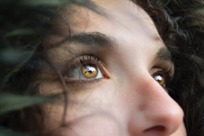
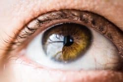
To understand what a corneal abrasion is first you must understand a little about the parts of the human eye.  The cornea is the clear, protective covering over the iris, which is the colored part of the eye, and the pupil — the black circle in the middle of the eye. The cornea is important for both vision and protection of the eye.  A corneal abrasion  then, is a scratch on that outermost covering of the eye.  Common causes include: being poked in the eye (makeup or finger), getting debris in the eye, aggressively rubbing the eye, bad hygiene with contacts, or potentially chemical burns.  If any of these events or circumstances have happened and you are experiencing pain and discomfort then you may have a corneal abrasion.  Symptoms that may also give evidence to this include:  tearing of the eye, feeling like something is in your eye, redness, eye pain, stinging or even blurred vision.

No one likes having something in their eye or even the feeling like something is in the eye.  One of the most important things to remember, should this happen to you, is NOT to rub the eye.  This could lead to the abrasion or cause more damage.  instead let the eye water to flush debris out, pull the top eyelid over the bottom eyelid and allow for watering or gently wash the eye with warm water.  If this does not rid your eye of the symptoms and pain see your doctor.

If your eye doctor believes you have suffered a corneal abrasion he/she may give you eye drops or ointment to prevent infection and to stop the pain.  In the case of a minor corneal abrasion the scratch will most likely heal on its own.  Larger or deeper scratches make take longer to heal and will require a patch, discontinued use of contacts and shaded glasses to shield the eye.

Eye Floaters

Do you experience small moving spots that appear in your field of vision?  Do you notice these “floaters”  more when there is bright light or the background is a solid color like a blue sky or a solid white wall?  Then you may be experiencing eye floaters.  While these shadows or spots may be annoying at times they rarely interfere with good vision.  Lets look at what these floaters are and what may be causing them.

What are Floaters?

According to the Mayo Clinic, Floaters are  spots in your vision. They may look to you like black or gray specks, strings or cobwebs that drift about when you move your eyes and appear to dart away when you try to look at them directly. Symptoms may include: spots of black or grayish blobs, spots that drift through your field of vision, spots that move when your eyes move, or possibly squiggly lines that are fairly transparent.

Causes of Floaters

Eye Floaters are usually caused by small flecks of a protein called collagen. The most common causes of these flecks of protein include:

  • Age-related eye changes- As we age the vitreous jelly that fills our eyeballs begins to liquify.  As the vitreous shrinks and sags, it clumps and gets stringy. Bits of this debris block some of the light passing through the eye, casting tiny shadows on your retina.
  • Inflammation – This swelling at the back of the eye called the uvea  may be causing the floaters to appear.
  • Bleeding in the eye. Bleeding into the vitreous can have many causes, including injury and blood vessel problems. Seek immediate medical attention of floaters appear after a blunt injury to the eye.
  • Torn Retina – Retinal tears can occur when a sagging vitreous tugs on the retina. Without treatment, retinal tear may lead to retinal detachment. Due to this more serious cause it is imperative to see a doctor if you are suddenly getting many more floaters than usual.  It is also important to immediately seek medical help if these floaters not only increase in numbers and frequency but also are accompanied by flashes of light.  Retinal tears are sight threatening conditions that should be treated immediately by an eye doctor.

What causes Strabismus and how can it be treated?

While holding or playing with your child, you notice that something is not quite right with the movement of his/her eyes.  Maybe one is looking in one direction and the other is off by a bit.  Or maybe you notice that when your child looks back and forth the eyes are not moving in coordination with each other.  What is this crossing of the eyes and how serious is it?  The first thing you should do is set up an appointment with your child’s pediatrician who will most likely do an eye exam and (depending upon the age of the child) recommend a visit to an ophthalmologist.  In the end the diagnosis could be strabismus.

What is strabismus?

Crossing of the eyes, or strabismus as it is medically termed, is a condition in which both eyes do not look at the same place at the same time. It occurs when an eye turns in, out, up or down and is usually caused by poor eye muscle control or a high amount of farsightedness. The disorder is very common in children, affecting four percent of children age 6 and younger. It does tend to run in families.  It is normal for children under 6 months of age to experience occasional crossed eyes because their brains are still developing the ability to see normally.  They will likely grow out of it. However in the case your child is being monitored for this and the symptoms do not disappear there are treatments available.

Treatment for Strabismus

Common treatments for these and other kinds of strabismus include special eye drops, eye patches, appropriate eyewear, vision therapy, and—in extreme cases—surgery. Most of the time, strabismus can be fixed if caught early enough and treated appropriately.

  • Eyewear – his method may help people who have crossed eyes due to an uncorrected farsightedness
  • Eye patches are commonly used to cover the stronger eye to build up the muscles in the weaker eye.  Eye drops to blur the vision of the stronger eye can also be used to get the same results.
  • Surgery – Eye muscle surgery may be needed if the eyes still do not move correctly. Different muscles in the eye will be made stronger or weaker.

Tips For Eye Emergencies

We know we don’t want to plan for emergencies to happen, but there’s nothing more important than being prepared in the case they do occur. Eye emergencies can happen anywhere. You could be at home, at work, at school, or doing chores.

Common Occurrences Of Emergency Situations

  • While spring cleaning, cleaning solution gets accidentally sprayed in your eye.
  • Your son is pitching the game of his life until the batter hits a ball directly at his face and his eye is completely swollen shut.
  • A day at the beach turns painful when the wind picks up and the sand gets lodged in your eye causing pain and blurred vision.

Eye Emergencies Can Include:

Eye emergencies range from cuts, scratches, objects in the eye and burns, all that way to chemical exposure, and blunt injuries to the eye or eyelid. An eye emergency, therefore, could be any event where a person’s eyesight is at risk.

It is extremely important to seek medical attention for any eye/eyelid injury or problem.

When To Seek Immediate Care:

Seek IMMEDIATE emergency care if you believe your eyesight is in jeopardy, if you are in severe pain, or if you have any of the following symptoms:

  • Sudden vision loss
  • Pain in or around the eye redness accompanied by pain in the eye.  This could be from an unknown cause or from a blunt injury to the eye area.
  • Halos (colored circles around lights)
  • New floaters (spots, strings, cobwebs, or shadows before the eyes)
  • Bulging of the eye or swelling of eye tissues.
  • Flashes or streaks of light
  • Double vision
  • Sudden crossed, turned or “wandering” eye
  • Bleeding of the eye. discharge, crusting or excessive tearing
  • Eyelids stuck together, especially upon awakening
  • Stinging or burning in the eye
  • The intense sensation of something in the eye
  • Pupils of unequal size
  • Headaches and strains

Tips from the US National Library of Medicine:

After calling the doctor or ambulance, depending upon the severity of the problem, you may want to consider the following tips from the U.S. National Library of Medicine.

Small Objects In The Eyes

Try not to itch or rub the eye.  Try gently rinsing the object out with water. Seek medical attention to remove the object whether it is large or small.

Chemical Exposure

Flush with cool tap water right away. Turn the person’s head so the injured eye is down and to the side. Holding the eyelid open, allow running water from the faucet to flush the eye for 15 minutes. If the person is wearing contact lenses have them remove them after the water rinse.  Seek medical help right away. Do not delay.

Blunt Force Injury To Eye

Apply a cold compress or eye pack to the area to bring down the swelling.  Seek medical attention to be sure the eye is not injured. 

Lazy Eye

Most new parents spend countless hours looking into the eyes of their newborn or toddler enjoying the delight or wonder they see in them.  Sometimes, however, parents notice something other than delight.   They may notice a wandering of one eye, or eyes that don’t appear to work together.  If this is noticed beyond the first few weeks of life, a check by your Primary Care physician may be in order.  He or she may refer your child to a specialist in eye conditions (ophthalmologist or optometrist). Lazy eye is one possibility for this “wandering” especially if the baby was premature, had low birth weight or it runs in your family. Let’s look at the causes and potential treatments for children diagnosed with Lazy Eye.

Main Causes Of Lazy Eye

There are three common causes of Lazy Eye.

  • Strabismic – this very common cause of lazy eye is caused by an imbalance in the muscles responsible for positioning the eyes.
  • Deprivation – this occurs if there is a problem with one eye, such as a cloudy area in the lens (cataract). This “deprives” the child of clear vision in the eye.
  • Refractive – This is the result of a significant difference in the vision in each eye.  This might be caused by nearsightedness, farsightedness or an imperfection on the surface of the eye (astigmatism).

Treatments For Lazy Eye

Ideally, a lazy eye will be diagnosed in childhood to increase the success of treatment.

  • If the condition is caused by significant differences in sight such as near or farsightedness in the two eyes then corrective lenses may be in order.
  • For children with one weak eye, a patch over the stronger eye may help build up the vision in the weaker one.  This is especially helpful in children over 4.
  • Eye drops to blur the vision of the stronger eye to help strengthen the weaker one are also available.  A daily or twice-weekly drop of a drug called atropine can temporarily blur vision in the stronger eye. This will encourage your child to use his or her weaker eye and offers an alternative to wearing a patch.
  • Surgery may be needed to repair eye muscles if your child has crossing or wandering eyes.  Your child may also need surgery if he or she has droopy eyelids or cataracts.

Are Your or Your Child Experiencing Trouble With Lazy Eye?

Contact BEPS for a consultation. We can discuss the best practices for getting both eyes as strong and healthy as possible. Reach out to us today!

Corneal Ulcers

If you have noticed a sore or opening on the clear outer covering of your eye(cornea), you may be experiencing a corneal ulcer.  Some ulcers may appear as a gray to white area on the normally transparent cornea. However, some corneal ulcers may be too small to see without adequate magnification and illumination. This can sometimes be caused by bacterial or viral infections or even overuse or misuse of contacts among other reasons.   Anyone with an irritated eye that does not improve quickly after removing a contact lens or after mild irrigation should contact an ophthalmologist immediately.

Here are some quick facts about corneal ulcers: how they form, their symptoms and treatments.

Possible Signs and Symptoms of a corneal ulcer –

  • Redness, pain, a feeling that something is in the eye.
  • Pus or thick discharge draining from the eye.
  • Blurriness or pain when looking at bright lights.
  • Swelling of the eyelid.

Causes – There are a wide variety of causes of corneal ulcers, including:

  • Bacterial infections can cause corneal ulcers and are common in people who wear contact lenses.
  • Viruses such as  herpes simplex virus (the virus that causes cold sores) and the Varicella virus may cause corneal ulcers.
  • Fungal infections can cause corneal ulcers and may occur with improper care of contact lenses or overuse of eye drops that contain steroids.

Treatment Options –

  • Anti-infective agents directed at the inciting microbial agent will be used in cases of corneal ulcer due to infection. (Drops, or possibly oral medications)
  • In cases aggravated by dryness or corneal exposure, tear substitutes will be used, possibly accompanied by patching or a bandage contact lens.
  • In corneal ulcers involving injury, the inciting agent must be removed from the eye- using a slit lamp microscope to to remove the particles causing the injury.
  • In extreme cases surgery may be needed to debride the ulcer.

What is a sty in the eye?

If you have ever had a small, painful lump along the edge of your eyelid then you have probably had a sty (also spelled stye). While styes are fairly common and are usually visible on the outer eyelid, they have formed deeper inside the eyelid in some cases as well.  Let’s look at the causes, types and treatments of styes to keep you informed in case you suspect that you may be suffering from one of these painful blockages.

What is a sty and what causes it?

A sty is an external bump on the eyelid that swells and then eventually bursts then heals.  The sty originally forms due to a blocked gland that needs to become cleared.  There are two main types of sty lumps.  These include: hordeolum and chalazion. Each has different causes and treatments.

Hordeolum

A hordeolum is a blockage of one of the sweat glands found in the skin of the lid and base of the eyelashes, or one of the small sebaceous glands found at the base of the eyelashes.

Chalazion

A chalazion is a blockage of a meibomian gland, which is a special sebaceous gland unique to the eyelids.

The sty can occur due to a gland becoming blocked with its own secretion or possibly from makeup, dust or other foreign substances.  It can also be caused by a sluggish gland that was infected or had some sort of trauma.

What are the treatments for a sty on the eyelid?

A non-infected hordeolum will resolve on its own. Warm compresses may help soften the material in the gland, easing the drainage of the gland’s contents. A non-infected chalazion similarly will resolve on its own, though over a much longer period of time. A small chalazion may resolve within weeks, while larger ones may resolve over the course of a year. The most conservative treatment is application of frequent warm compresses. Steroids can be injected into the lesion, often resulting in a speedier resolution. However this carries a small risk of bleeding/bruising, depigmentation/thinning of the skin, scar, pain, and in very rare cases, loss of vision. Finally, the chalazion can be incised and drained. This is the most invasive method and is reserved as a last resort by most eye doctors.

What is Conjunctivitis?

If you have ever woken up with itchy eyes accompanied by a thick, crusty discharge or green or yellow mucus coming from your eye then you may have the dreaded
“pink eye” or conjunctivitis.  Other symptoms that are experienced with conjunctivitis include:  redness in the whites of the eyes or the inner eyelid, blurriness, burning or even sensitivity to light.  If you are suffering from any of these symptoms then consult your primary care physician for a swab of the effected area.  While this disease is not a serious health risk it should be dealt with quickly through your doctors office.

What is conjunctivitis?

Conjunctivitis, also known as pinkeye, is an inflammation of the conjunctiva. The conjunctiva is the thin clear tissue that lies over the white part of the eye and lines the inside of the eyelid.

What causes pink eye?

This eye disease has several possible causes such as: a virus, a bacterial infection such as gonorrhea or chlamydia, irritants such as shampoos, dirt, smoke, and pool chlorine
or possibly allergies, like dust, pollen, or a special type of allergy that affects some contact lens wearers.

What treatments are available for pink eye?

The treatment proscribed by your doctor will depend mostly upon the cause of the disease.  If bacteria was the root cause then antibiotics, in the form of eye drops, ointments, or pills will be the treatment.  If a virus is the cause (such as the virus that causes the common cold) then it must run its course over the next 7-10 days.  Warm compresses can help with the discharge.  Throw away all contacts and makeup that you used during the period before being diagnosed.  If irritants or allergies are the cause of the discharge then washing out eyes and removing irritants from laundry, home or other areas that will affect your eyes.

What Is Astigmatism?

Are you or a loved one suffering from the following symptoms- distortion or blurring of images at all distances, headaches, and fatigue or even squinting and eye discomfort or irritation? These symptoms may not necessarily mean a diagnosis of astigmatism, however, if you experience one or more of these symptoms, contact your ophthalmologist for a complete exam. Let’s look at the causes and treatment for astigmatism.

What Is Astigmatism?

What Is Astigmatism?

Astigmatism is an imperfection in the curvature of your cornea — the clear, round dome covering the eye’s iris and pupil — or in the shape of the eye’s lens. Astigmatism is very common. In most cases, people with astigmatism are born with this condition. The reason why corneal shape differs from person to person is unknown, but the likelihood of developing astigmatism is inherited.

Treatment For Astigmatism

Treatment For Astigmatism

Depending upon the degree of astigmatism will determine the treatment needed. If the degree of astigmatism is slight and there are no other problems of refraction, such as nearsightedness or farsightedness, corrective lenses may not be needed.

Eye Care in Boston

If the degree of astigmatism is great enough to cause eye strain, headache, or distortion of vision, corrective lenses will be needed for clear and comfortable vision. Your ophthalmologist will perform precise tests during your exam to determine the ideal lens prescription. Refractive surgery also may be an option for correcting some forms of astigmatism.

What is Age Related Macular Degeneration?

Age Related Macular Degeneration (AMD) is a common eye condition and a leading cause of vision loss among people age 50 and older. The eye’s macula (small part of the retina) deteriorates and fine details in central vision become difficult to see. Many older people develop macular degeneration as part of the body’s natural aging process. There are different kinds of macular problems, but the most common is age-related macular degeneration.

Symptoms of AMD – Blurriness, dark areas or distortion in your central vision, and perhaps permanent loss of your central vision. It usually does not affect your side, or peripheral vision.

Who is at risk?  Age is a major risk factor for AMD. The disease is most likely to occur after age 60, but it can occur earlier. Other risk factors for AMD include:

  • Smoking. Research shows that smoking doubles the risk of AMD.
  • Race. AMD is more common among Caucasians than among African-Americans or Hispanics/Latinos.
  • Family history. People with a family history of AMD are at higher risk.

Causes of AMD – include the formation of deposits called drusen under the retina, and in some cases, the growth of abnormal blood vessels under the retina. With or without treatment, macular degeneration alone almost never causes total blindness. People with more advanced cases of macular degeneration continue to have useful vision using their side, or peripheral vision.

The early and intermediate stages of AMD usually start without symptoms. Only a comprehensive dilated eye exam can detect AMD. Consult with your eye doctor is you have any symptoms of macular degeneration.